

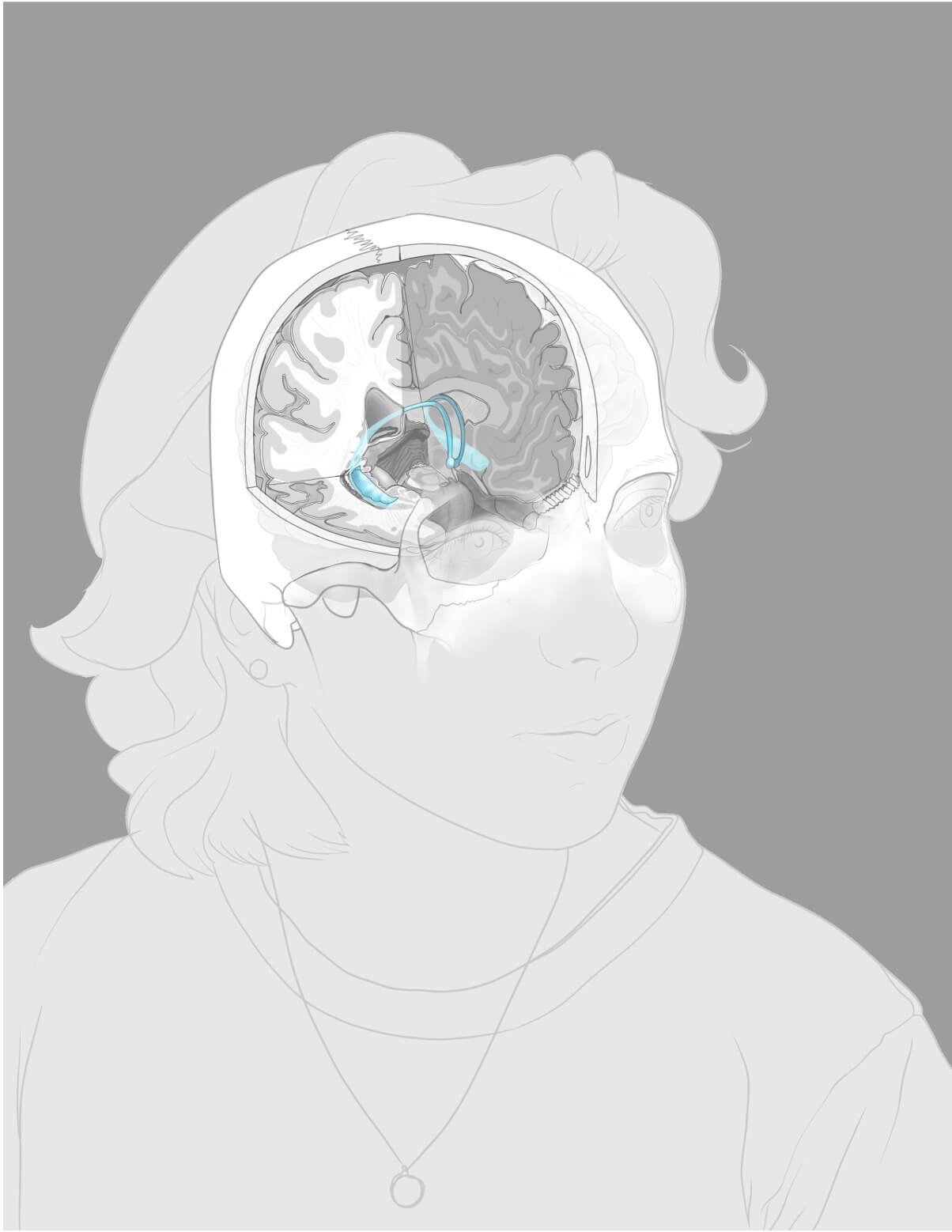
Neuroanatomy Self-Portrait: Vasculature of the Hippocampus
Purpose
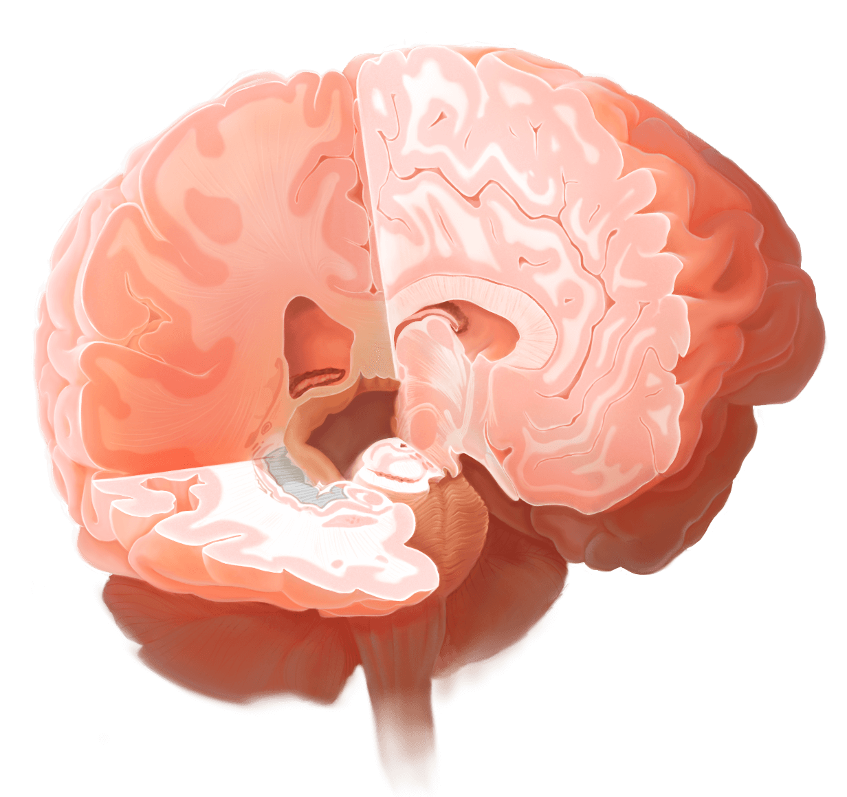
The main goals of this poster are to depict the 3D structure of the hippocampus relative to brain cross-sections, and to visualize the 3D structures of hippocampal vasculature. I was inspired to make this piece because, as a student, I struggled to understand the hippocampus’s position in the brain when looking at CT data. I thought showing the hippocampus in situ in cross sections would help other anatomy students.
Audience
Client
Content advisor
Undergraduate anatomy students
Intended format
Tools used
Science poster
3DSlicer, Zbrush, Autodesk Maya, Adobe Photoshop, Procreate, Adobe Illustrator
Shelley Wall, University of Toronto
Kristy Cheung, University of Toronto
Portrait reference
This project is a self portrait. Many photos were taken to allow for different angles for visualizing the brain. Photography credit to Julia Russo.


Brain maquette
After considering multiple sources, I downloaded a 3D brain file cleaned from CT data from NIH. I then uploaded the OBJ file into Maya to visualize it. I also downloaded this brainstem and cerebellum, as those structures were not very clear in the brain file.
To visualize relative locations of the skin, cranium, brain, and hippocampus, I sectioned the Colin 27 CT scan from the McGill University in 3D slicer, and imported the separate layers into Maya.
I overlayed the CT data and the cleaned brain assets on top of each other, to understand relative locations. I then used Maya to create a mock hippocampus over the approximate location from the CT data.


Brain cross-section
It was important for me to make sure my cross-sectional cuts were in perspective and accurate. To align my planes, I used illustrations from Nieuwenhuys et al. (2016), and the orientation guide to ensure that the cuts on the model matched those from the textbook.
I chose to cut coronal, sagittal, and axial sections, as I found it difficult to understand the location of the hippocampus in medical imagery, so I thought it would be helpful to visualize its 3D form within all three planes. I chose to make my axial section perpendicular to the brainstem, so that the cut would lie flush with the inferior surface of the hippocampus.

Brain cross-section
Using my maquette and my portrait photograph as references, I created my cross-section illustration in Procreate. I also referenced real and plastic skulls supplied by BMC, as well as skull 3D models from the Neurosurgical Atlas and Elsevier’s complete Anatomy.
I chose to cut coronal, sagittal, and axial sections, as I found it difficult to understand the location of the hippocampus in medical imagery, so I thought it would be helpful to visualize its 3D form within all three planes. I chose to make my axial section perpendicular to the brainstem, so that the cut would lie flush with the inferior surface of the hippocampus.

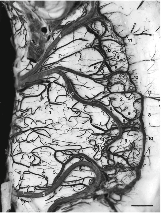
Inset Research
When researching the blood supply to the hippocampus, I found the structural organization very interesting, but difficult to wrap my head around. I thought that rather than a textual description, the spatial relationships between the structures was best tackled almost purely visually.
I decided to create visuals of blood supply to the hippocampus in general, as well as 3D cross sectional views to understand the spiral pattern of the vessels within the hippocampus itself.
•Due to the high number of different blood vessels associated with the hippocampus, I decided to remove explanatory text and only use labels. I was also inspired by science posters, which serve both an aesthetic and didactic purpose.
Images from Duverney et al. (2013).


Comprehensive draft and Layout
Inspired by the spiral shape of the hippocampus, I decided to use the Fibonacci sequence to lay out my inset!


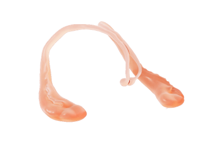
Inset Maquettes
Referencing Duverney et al. (2013), I sculpted a hippocampus from scratch using Zbrush, and extruded an SVG line drawing into a cut section using Adobe Illustrator and Maya.
I then added the Arnold standard surface skin preset and adjusted it to a material I was content with, then rendered the maquette to use as the base for my illustrations.
I used an 18mm camera view to have some distortion, as I wanted the inset to appear to be diving towards the viewer.


Inset Rendering
I used diagrammatic and visualizations of vasculature from Duverney et al. (2013) to achieve a balance between a flat diagram and reality
I used opacity masks, alpha lock, and drop shadows to convey how the vessels rest on top or dive into the cross sections, and are affected by the light.


Brain Rendering
I rendered my sculpted hippocampus inside the brain, using my previous sectioned CT data to place it accurately.
I used an adjusted skin material preset from Maya, and then rendered both the brain and hippocampus.
I rendered the hippocampus multiple times with a realistic texture as well as a glow, to allow flexibility in the final render.
I further edited colours and shadows using adjustment layers and masking in Photoshop.
Starting with my rendered maquette, I used the smudge tool to approximately match the maquette to my comprehensive draft, then cleaned up the linework in Procreate.
I then used a combination of soft/hard light and multiply layers to gradually build in depth and form.


Final Version
I finished by rendering the portrait and adding labelling. Overall, I am proud of how this piece evolved and turned out.
I am especially pleased with how the insets turned out, I think that spending time with my maquettes helped front load my workflow.

Academic references
Crossman, A. R., & Neary, D. (2024). Neuroanatomy: An Illustrated Colour Text (6th edition). Elsevier Canada.
Duvernoy, H. M., Cattin, F., & Risold, P.-Y. (2013). The Human Hippocampus: Functional Anatomy, Vascularization and Serial Sections with MRI (4th ed. 2013 edition). Springer.
Gerardin, E. (2012). Morphometry of the human hippocampus from MRI and conventional MRI high field.
Johnson, A. C. (2023). Hippocampal Vascular Supply and Its Role in Vascular Cognitive Impairment. Stroke, 54(3), 673–685. https://doi.org/10.1161/STROKEAHA.122.038263
Kim, G. H., Lee, J. H., Seo, S. W., Kim, J. H., Seong, J.-K., Ye, B. S., Cho, H., Noh, Y., Kim, H. J., Yoon, C. W., Oh, S. J., Kim, J. S., Choe, Y. S., Lee, K. H., Kim, S. T., Hwang, J. W., Jeong, J. H., & Na, D. L. (2015). Hippocampal volume and shape in pure subcortical vascular dementia. Neurobiology of Aging, 36(1), 485–491. https://doi.org/10.1016/j.neurobiolaging.2014.08.009
Nieuwenhuys, R., Voogd, J., & Huijzen, C. van. (2016). The Human Central Nervous System: A Synopsis and Atlas (4th edition). Springer Nature.
Perosa, V., Priester, A., Ziegler, G., Cardenas-Blanco, A., Dobisch, L., Spallazzi, M., Assmann, A., Maass, A., Speck, O., Oltmer, J., Heinze, H.-J., Schreiber, S., & Düzel, E. (2020). Hippocampal vascular reserve associated with cognitive performance and hippocampal volume. Brain, 143(2), 622–634. https://doi.org/10.1093/brain/awz383
Ribas, G. C. (2010). The cerebral sulci and gyri. Neurosurgical Focus, 28(2), E2. https://doi.org/10.3171/2009.11.FOCUS09245
What is vascular dementia? | Alzheimer’s Society. (n.d.). Retrieved October 6, 2024, from https://www.alzheimers.org.uk/about-dementia/types-dementia/vascular-dementia
Who gets vascular dementia? | Alzheimer’s Society. (2022, June 21). https://www.alzheimers.org.uk/about-dementia/types-dementia/risk-factors-vascular-dementia
Yang, X., Chen, C., Wang, A., Li, C., & Cheng, G. (2023). Imaging, Genetic, and Pathological Features of Vascular Dementia. European Neurology, 86(4), 277–284. https://doi.org/10.1159/000531088
Interested in working together? Let’s talk about it.
I work on projects big and small. Let me know what you have in mind and we can chat details.